


A man has been spared a major brain operation after surgeons in Leeds performed a pioneering UK-first procedure, displacing his eye to treat a life-threatening brain aneurysm.
Andrew Wood was “shocked” to discover he had the condition – a swelling in a blood vessel that can prove fatal if it ruptures.
Traditionally, such a diagnosis would necessitate a craniotomy, where a section of the skull is removed and the brain moved to access the affected area.
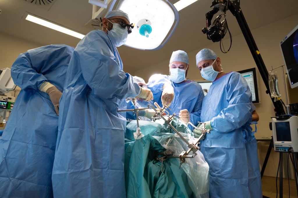
However, experts at Leeds Teaching Hospitals NHS Trust successfully employed an innovative keyhole technique, accessing the aneurysm directly through Mr Wood’s eye socket.
This marks the first time this groundbreaking operation has been carried out in the United Kingdom.
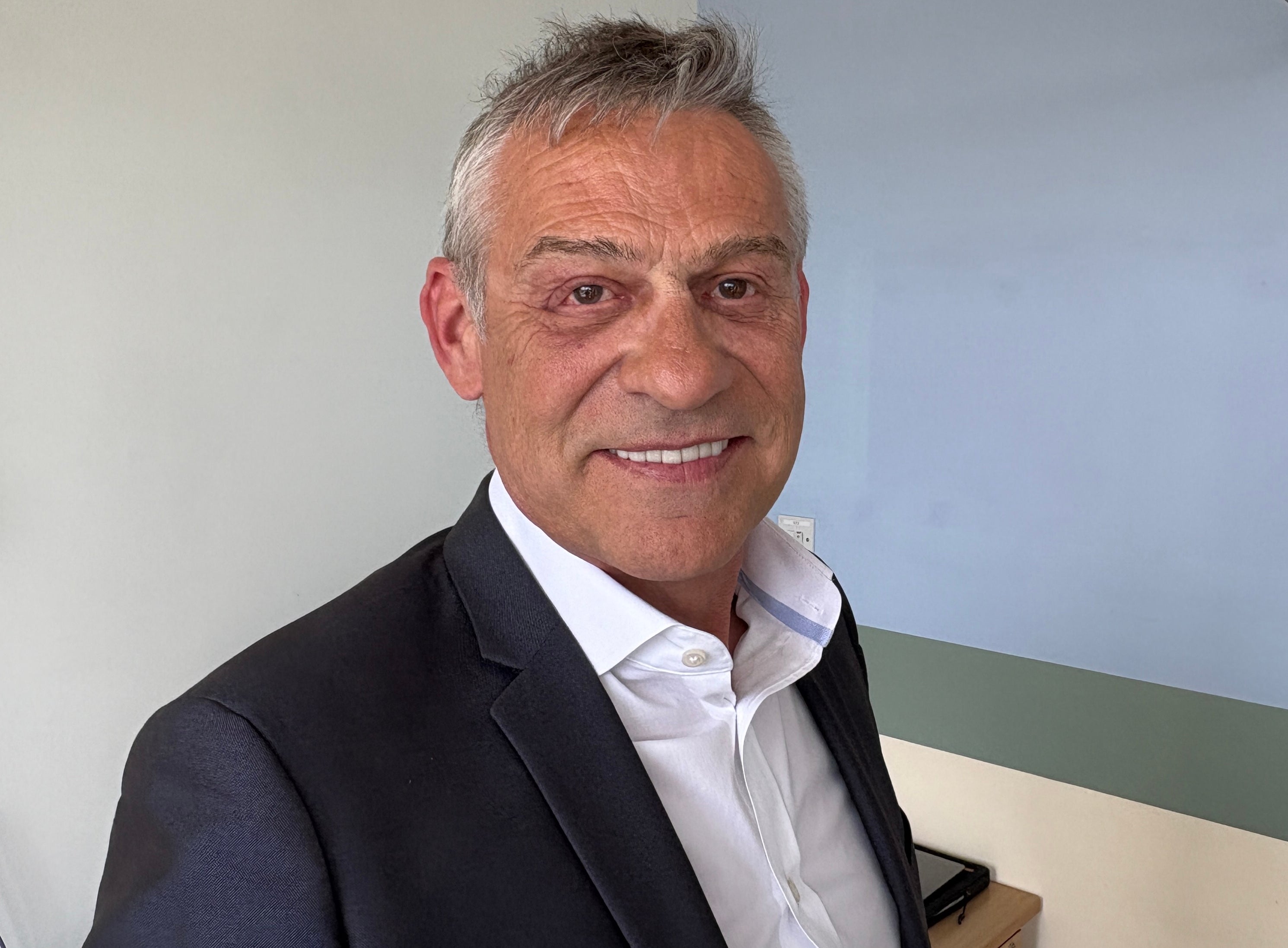
“This meant that we could directly access the aneurysm without even having to touch the brain,” said consultant neurosurgeon Asim Sheikh.
Mr Wood was in hospital for just one night after the procedure, when traditionally patients would be in hospital for around a week.
The 61-year-old grandfather, who works as a builder, returned to work in May just a few weeks after the pioneering operation in February.
The aneurysm was found while Mr Wood was being scanned for a separate medical issue last spring.
“I was shocked. I didn’t have any symptoms whatsoever,” he told the Press Association.
“Mr Sheikh got his hands on my case and it just went 100 miles an hour from there.
“I’m in the building trade so the way it was explained to me was: you can do something causing minimum damage and get the same result.
“I thought it was great.”
One treatment is clipping, where a neurosurgeon carefully places a tiny metal clip across the base of the aneurysm to prevent it from rupturing.
The team of experts from Leeds used the new minimally invasive technique to get to Mr Wood’s aneurysm.
Surgeons made a “tiny” incision at the side of Mr Wood’s eye and a cut on the outer wall of the eye socket to gain access to the aneurysm.
Mr Sheikh said that Mr Wood “got the best of both worlds” by having the surgery.
The team behind the procedure performed a similar operation in 2024 when they removed a brain tumour through a patient’s eye socket – another UK-first.
Mr Sheikh told the Press Association: “As we gained more experience, we realised that the same approach could be used to access other pathologies in the similar area – in Andrew’s case a brain aneurysm.
“His aneurysm happened to be in a very easily accessible area, which would have been perfect for this technique.
“And that means he can get the best of both worlds of the surgical treatment of his aneurysm – the best possible, durable cure for his aneurysm while cutting down on the drawbacks of having surgery including big cuts and scars, big incisions on the head and also the morbidity of going through the brain and retracting the brain – all that is completely taken away by this minimal access surgery.”
To prepare for the procedure, biomechanical engineers from the hospital created a bespoke 3D printed model of Mr Wood’s eye socket and surrounding skull base anatomy as well as the aneurysm.
This allowed Mr Sheikh and consultant maxillofacial surgeon Jiten Parmar to prepare and rehearse the procedure, tailored specifically to Mr Wood’s anatomy.
Mr Sheikh said the operation is a “significant step forward in minimally invasive brain surgery in the UK”.
He added: “It was easily accessible through this approach.
“Traditionally, we would have done a traditional craniotomy, which means making a big opening in the skull and reaching it through that to where his aneurysm was situated between two parts of the brain called the frontal and temporal.
“So traditionally, you have to split the two halves, pull them apart to get to the aneurysm, and then place a metal clip across it.
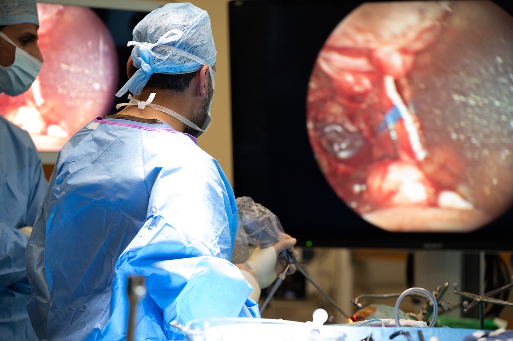
“With this technique, we didn’t even have to touch the brain, we didn’t have to put any retraction on the brain and we were directly approaching the aneurysm, which meant that the morbidity of the operation itself was substantially reduced.”
Experts at the hospital also 3D-printed custom-made retractors to protect the eye during the procedure.
“That meant we weren’t pushing on the on the eyeball as well. And that created the corridor which allowed us to access the aneurysm,” Mr Sheikh said.
Mr Parmar added: “This case highlights how working in partnership across specialities, combining decades of experience and working closely with our engineering team can result in a better outcome for our patients.
“Most importantly, we achieved a brilliant result for the patient who went home the next day.”
Mr Wood added: “I was in hospital for just one night.
“I was asked to make some toast and a cup of tea to make sure my faculties were OK, I had another quick scan and then they asked if I wanted to go home.
“It has been perfect since, I’ve had no double vision, no pain, I’m back at work – just have to watch out that I don’t hit myself with a two by four.”
He went on: “Thanks to the skill of the surgical team, I’ve been given a second chance.
“I’m really grateful for their kindness and expertise – they are an outstanding team. I’m proud to have been part of such an important procedure and I’m amazed at how quickly I recovered and returned home.”
-and-Peter-Johnstone-in-1987.png?trim=34,0,36,0&width=1200&height=800&crop=1200:800 "‘I investigated one of Britain’s most notorious missing person cases – I never gave up’ – UK Times")

